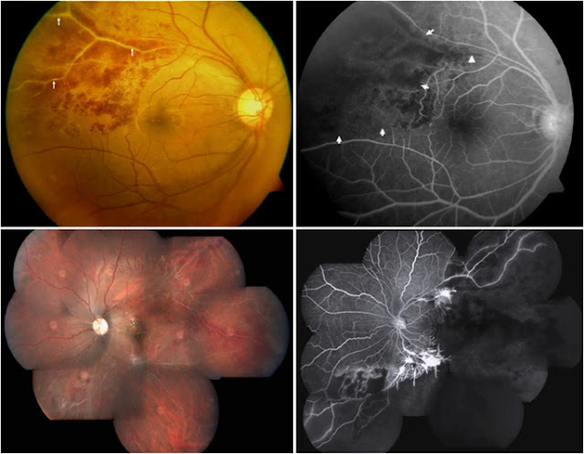
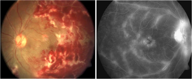
Eales Disease is a rare disorder of sight that appears as an inflammation and white haze around the outercoat of the veins in the retina. The disorder is most prevalent among young males and normally affects both eyes. Usually, vision is suddenly blurred because the clear jelly that fills the eyeball behind the lens of the eye seeps out (vitreous hemorrhaging).
 |
| Eales' disease |
Eales Disease usually presents as blurred vision resulting from oozing of the clear jelly-like substance from behind the lens of the eye. At the onset of the disorder, the small outer veins of the retina show sheathing (encapsulation or covering). As the disease progresses, the inflammation around the veins in the retina extends further behind the lens. Eales Disease may also be associated with peripheral retinal neovascularization which is the formation of new blood vessels on the outer part of the retina.
📖 The Massachusetts Eye and Ear Infirmary Illustrated Manual of Ophthalmology 5th EditionThe more advanced cases of Eales Disease are characterized by a non- inflammatory degenerative disease of the retina (retinopathy) and extensive bleeding in the retina. The colorless jelly that fills the eyeball behind the lens oozes from the retina (vitreous hemorrhage) and, in rare cases, the retina may become detached. A reddish discoloration of the iris may be present (rubeosis iridis), and there may be loss of vision and damage to the optic disk (neovascular glaucoma). Clouding of the lens of the eye that obstructs the passage of light (cataracts) may develop as the disease progresses.
The exact case of Eales Disease is not known. This disorder seems to occur spontaneously because no precipitating factors such as injury, infection, or heredity appear to be involved.
Symptoms of the following disorders can be similar to those of Eales Disease. Comparisons may be useful for a differential diagnosis. Arteriosclerotic Retinopathy alludes to a series of changes in the retina caused by hardening of the arteries (arteriosclerosis) serving the retina. The characteristics of this disorder are bleeding in the retina, thick fluid oozing from the retina, impaired oxygenation of the retina, and hardening of the walls of the vision impairment.
Treatment of Eales Disease is symptomatic and supportive. The surgical process of coagulating tissue with a laser beam (laser panretinal photocoagulation) may be used to eliminate the deficiency of blood in the retina caused by constriction of blood vessels and to slow down excessive formation of blood vessel tissue.
Hemorrhaging of the clear jelly that is behind the lens of the eye (vitreous) and detachment of the retina) may be helped by the removal of the dark pigmented disk and jelly-like substance behind the retina (pars plana vitrectomy.
Henry Eales (1852 - 1913), British ophthalmologist.
 |
| Henry Eales |
Henry Eales was born at Newton Abbot, the son of the vicar of Yealmpton in Devonshire. While an apprentice to the village doctor, and following an outbreak of scarlet fever which led him to test patient’s urine for the presence of protein, he incidentally examined his own and found himself to have heavy proteinuria. As a result he had a year’s convalescence before he enrolled in medicine at the University College, London.
Eales had a fine undergraduate record and graduated M.R.C.S. in 1873 and then interned at the Birmingham and Midland Eye Hospital. He was demonstrator in anatomy and medical tutor at Queen’s College, and in 1878 was appointed honorary surgeon to the Eye Hospitals, where he remained for 35 years. He was well known for his abilities with the ophthalmoscope and built up a very big consulting practice. He wrote a number of papers, amongst which was a review of the appearance of the retina in patients with renal disease.
Apart from occasional migraine he enjoyed good health and the proteinuria did not return. Shortly before his death he developed pain in his left calf which waxed and waned for 10 days, forcing him to go to bed, and he died sometime thereafter following syncopal attack, possibly due to pulmonary embolus.
References
1
Eales H . Retinal haemorrhages associated with epistaxis and constipation. Brim Med Rev 1980; 9: 262.
2
Eales H . Primary retinal haemorrhage in young men. Ophthalmic Rev 1882; 1: 41.
3
Wardsworth. Recurrent retinal haemorrhage followed by the development of blood vessels in the vitreous. Ophthalmic Rev 1887; 6: 289.
4
Elliot AJ . Recurrent intraocular haemorrhage in young adults: Eales’ disease. A preliminary report. Trans Cand Ophthalmol Soc 1948; 46: 39.
5
Kimura SJ, Carriker FR, Hogen MJ . Retinal vasculitis with intraocular haemorrhage. Classification and results of special studies. Arch Ophthalmol 1956; 56: 361.
6
Keith-Lyle T, Wybar K . Retinal vasculitis. Br J Ophthalmol 1961; 45: 778.
7
Cross AC . Vasculitis retinae. Trans Ophthalmol Soc UK 1963; 83: 133.
8
Puttamma ST . Varied fundus picture of central retinal vasculitis. Trans Asia Pacific Acad Ophthalmol 1970; 3: 520.
9
Duke-Elder S, Dobree JH . System of Ophthalmology. Vol X. London: Henry Kimpton.
10
Murthy KR, Abraham C, Baig SM, Badrinath SS . Eales’ disease. Proc All lnd Ophthalmol Soc 1977; 33: 323.
Tags: eales' disease tuberculosis, eales' disease stages, eales' disease ppt, eales' disease tb, eales' disease treatment in india, eales' disease pdf, eales disease treatment, eales disease symptoms, eales disease, eales disease aao, eales disease and tuberculosis, eales disease article, eales disease fluorescein angiography, eales disease treatment in ayurveda, eales disease bilateral, eales disease causes, is eales disease curable, eales disease case report, eales disease clinical features, eales disease complications, eales disease cure, what is eales disease, eales disease differential diagnosis, eales disease dos times, best disease differential diagnosis, what causes eales disease, eales disease eye, does stargardt disease lead to blindness, eales disease fundus, best disease fundus, eales disease hereditary, eales disease in hindi, what disease are hereditary, is stargardt's disease hereditary, eales disease icd 10, eales disease investigations, eales disease images, eales disease ijo, eales disease mnemonic, eales disease ncbi, eales disease ophthalmology, eales disease ocular, eales disease review of optometry, eales disease pronunciation, eales disease prognosis, eales disease pubmed, eales disease prednisone, eales disease retina, eales disease review, eales disease radiology, eales disease slideshare, eales disease therapy, eales disease uptodate, eales disease uveitis, eales disease wikipedia, eales disease eye wiki


No comments:
Post a Comment