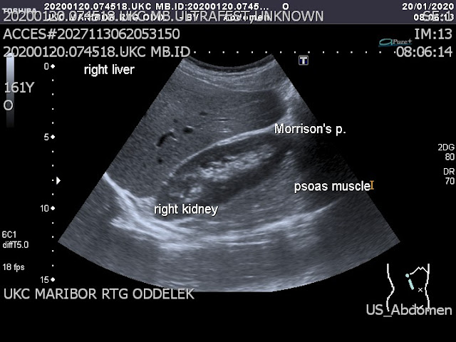
Morison’s pouch is also known as the posterior right subhepatic space or hepatorenal fossa. It separates the liver from the right kidney and is not filled with any fluid under normal conditions. It is a potential space, meaning a space that can occur between two adjacent structures that are normally pressed together. The anterior boundary consists of the right hepatic lobe and gallbladder.
 |
| Morison’s pouch |
Posteriorly, there is the right kidney, right adrenal gland, the second part of the duodenum, hepatic flexure, and pancreatic head. The transverse mesocolon lies inferiorly. The posterior right subhepatic space communicates with the right subphrenic space and right paracolic gutter.
📖 Gray’s Atlas of Anatomy 3th Edition
Probe position in assessment of hepatorenal recess – Morison's pouch
Fluid can collect in this space in different circumstances, such as ascites or hemoperitoneum. This fluid may be seen as an anechogenic border between the liver and the kidney on the ultrasound. If there has been recent trauma or bleeding in the abdomen, it can often be discovered in this location.
 |
| Ultrasound image of Morison's pouch |
Free fluid in the pouch other than bleeding is known as ascites. It can commonly be caused by heart, liver, or kidney conditions. Early visualization of fluid in the hepatorenal recess on the FAST scan may be an indication for urgent laparotomy.
Ultrasound examination
The liver, right kidney, and Morrison’s pouch are assessed in this view.
 |
| Free fluid in Morison’s pouch |
A transducer is put in the mid-axillary line at the lever of the 10th rib, with the marker on the probe pointing towards the patient’s head. Ideally, the kidney, liver, and diaphragm are seen at the same time. The hepatorenal recess should be placed in the center of the ultrasound screen. At this point, the probe is tilted around to assess Morrison’s pouch, liver, and kidney. It may be necessary to move one intercostal space inferiorly to evaluate the liver tip. Ribs may get in the way of a clear picture. In this case, the probe can be rotated around its axis, with the marker pointing slightly posteriorly (to get in between the ribs). Another way to avoid rib shadows is to ask the patient to inhale.
James Rutherford Morison (1853 - 1939). British surgeon.
 |
| James Rutherford Morison |
The treatment of infected suppurating war wounds. The Lancet, 1916, 2: 268-272. Introduction of "Bipp" in the treatment of wounds.
References
1: Nassar S, Menias CO, Nada A, Blair KJ, Shaaban AM, Mellnick VM, Gaballah AH,
Lubner MG, Baiomy A, Rohren SA, Elsayes KM. Morison's pouch: anatomical review
and evaluation of pathologies and disease spread on cross-sectional imaging.
Abdom Radiol (NY). 2020 Aug;45(8):2315-2326. doi: 10.1007/s00261-020-02597-1.
PMID: 32529262.
2: Meyers MA. Morison pouch. Radiology. 1995 May;195(2):578. doi:
10.1148/radiology.195.2.7724790. PMID: 7724790.
3: Veerapong J, Solomon H, Helm CW. Morison's Pouch peritonectomy in
cytoreductive surgery. Gynecol Oncol. 2013 Oct;131(1):214. doi:
10.1016/j.ygyno.2013.07.083. Epub 2013 Jul 14. PMID: 23863360.
4: Aguilera-Bohórquez B, Cantor E, Ramos-Cardozo O, Pachón-Vásquez M.
Intraoperative Monitoring and Intra-abdominal Fluid Extravasation During Hip
Arthroscopy. Arthroscopy. 2020 Jan;36(1):139-147. doi:
10.1016/j.arthro.2019.07.031. PMID: 31864567.
5: Brill PW, Olson SR, Winchester P. Neonatal necrotizing enterocolitis: air in
Morison pouch. Radiology. 1990 Feb;174(2):469-71. doi:
10.1148/radiology.174.2.2296656. PMID: 2296656.
6: Aguilera-Bohórquez B, Ramirez S, Cantor E, Sanchez M, Brugiatti M, Cardozo O,
Pachón-Vásquez M. Intra-abdominal Fluid Extravasation: Is Endoscopic Deep
Gluteal Space Exploration a Risk Factor? Orthop J Sports Med. 2020 Aug
4;8(8):2325967120940958. doi: 10.1177/2325967120940958. PMID: 32821761; PMCID:
PMC7412916.
7: Moore C, Todd WM, O'Brien E, Lin H. Free fluid in Morison's pouch on bedside
ultrasound predicts need for operative intervention in suspected ectopic
pregnancy. Acad Emerg Med. 2007 Aug;14(8):755-8. doi: 10.1197/j.aem.2007.04.010.
Epub 2007 Jun 6. PMID: 17554008.
8: Sharma A, Bhattarai P, Sharma A. eFAST for the diagnosis of a perioperative
complication during percutaneous nephrolithotomy. Crit Ultrasound J. 2018 Apr
3;10(1):7. doi: 10.1186/s13089-018-0088-1. PMID: 29616352; PMCID: PMC5882478.
9: Gilliam JW Jr, Schein CJ. The Morison pouch. Arch Surg. 1976
Mar;111(3):227-8. doi: 10.1001/archsurg.1976.01360210021003. PMID: 816330.
10: O'Connor G, Ramiah V, Breslin T, McInerney JJ, Brazil E. Looking beyond
Morison's pouch in focused assessment with sonography for trauma: penetrating
hepatobiliary trauma and a new sign for emergency physicians. Emerg Med J. 2013
Sep;30(9):778-9. doi: 10.1136/emermed-2012-201336. Epub 2012 Apr 4. PMID:
22492124.
morison’s pouch is anterior to which organ, morison pouch boundaries, morison pouch anatomy, morison pouch fluid ultrasound, morison pouch ultrasound, morison pouch radiology, morison pouch adalah, morison pouch ct, morison pouch, morison pouch ascites, morison pouch air, aszites morrison pouch, morison pouch collection, fluid in morison's pouch causes, morison pouch definition, morison pouch doccheck, what is morison pouch, where is morison pouch located, definition pouch, morison's pouch, morrison’s pouch, morrison's pouch, morison's pouch fluid radiology, fast morison pouch, hepato morison pouch, morison pouch intraperitoneal, morison's pouch is located in the, morison's pouch ct, morison koller pouch, morison's pouch location, what is morison's pouch, pouch of morison, morison pouch sign x ray, rutherford morison pouch, morrison pouch raum, morison pouch sign, morison pouch sono, morison pouch treatment, morison pouch tumor, best way to get rid of pouch, morison pouch usg, morison pouch ultraschall, morison pouch wiki


No comments:
Post a Comment