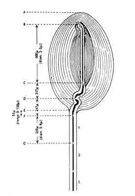
The structure of the Pacinian corpuscle was described by Pacini (1835). It is widely distributed in mammals and is similar to the Herbst corpuscles found in birds. The Pacinian corpuscle is an ovoid structure about 1 mm in length and is easily seen by the naked eye in a number of locations such as the mesentery.
 |
| Pacini's corpuscles |
On microscopic examination, the lamellar structure of the corpuscle is evident, the lamellae giving an appearance which has been likened to a section through an onion. The corpuscle is innerv!lted by a myelinated sensory axon of medium diameter which terminates within the center of the corpuscle.
📖 Clinical Neuroanatomy, 29th Edition
There it loses its myelin and terminates as an unmyelinated axon· Pacinian corpuscles are highly sensitive mechanoreceptors which respond only to rapid mechanical changes. They are particularly responsive to vibration and appear to subserve the type of sensibility known as vibration sense in man. The corpuscle has been extensively studied as to its morphology, its functional characteristics, and its projection to the central nervous system. The discrete nature and large size of this receptor have made it particularly attractive for the study of receptor mechanisms.
 |
| Diagram of a Pacinian corpuscle. |
The length of the corpuscle ranges from about 0.5 to 1.5 mm, with an average length of approximately 1 mm; the average width is about 0.7 mm. Occasionally, much smaller corpuscles are seen (Quilliam and Sato, 1955.) A single myelinated nerve fiber enters the ellipsoid corpuscle at one end; its course in the corpuscle for the first one-fourth of its length is tortuous and it retains its myelination over this length (Fig. 1). Following this, it enters the central core of the corpuscle and becomes thinner and unmyelinated. It travels along the longitudinal axis of the corpuscle toward the other end of the central core, where it frequently bifurcates. The diameter of the myelinated fiber just outside the corpuscle varies from about 4 to 7 jLm. One node of Ranvier lies within the corpuscle; the second node lies between 0 and 150 jLm from the central end of the corpuscle. The myelinated fiber in its intracorpuscular course has about the same diameter as immediately outside the corpuscle, although the thickness of the myelin tends to vary more during its course within the corpuscle.
Filippo Pacini (1812-1883), Italian antomist.
 |
| Filippo Pacini |
Pacini saw the corpuscles that are now named for him early in his career; indeed, he discovered them in a hand that he was dissecting as a student in the anatomy class in Pistoia hospital in 1831, when he was nineteen. He first saw the corpuscles around the digital branches of the median nerve, and suggested that they were "nervous ganglia of touch"; but he soon found them also in the abdominal cavity. Although he studied these corpuscles microscopically from 1833 on, Pacini published his research only in 1840, when his Nuovi organi scoperti nel corpo umano appeared. However, he had mentioned them at a scientific meeting in Florence in 1835.
The name "Pacini's corpuscles" was proposed in 1844 by Henle, and by Kölliker, who had confirmed their existence. In 1862, however, the Viennese anatomist Carl Langer claimed priority for Abraham Vater - although Vater's work, published in 1741, had been forgotten and was certainly unknown to Pacini. Vater noted the corpuscles in the skin of the fingers. He called them the papillae nervae and they were later depicted by Lehmann in 1741. They were apparently forgotten until they were rediscovered in 1831 by Pacini. At all events, Pacini was the first to describe the distribution of the corpuscles in the body, their microscopic structure, and their nerve connections; he also interpreted the function of the corpuscles as being concerned with the sensation of touch and deep pressure. In 1844 Friedrich Gustav Jacob Henle (1809-1885) and Albert Kölliker (1817-1905) named these structures Pacinian corpuscles.
References
No comments:
Post a Comment