Under normal circumstances, the tongue is a pink, muscular organ located within the oral cavity proper. It is kept moist by the products of the major and minor salivary glands, which aids the organ as it facilitates deglutition, speech, and gustatory perception. While there is significant variability in the length of the tongue among individuals, on average, the organ is roughly 10 cm long. It has three main parts:
 |
| Tongue |
The tip or apex of the tongue is the most anterior, and most mobile aspect of the organ.
The tip is followed by the body of the tongue. It has a rough dorsal (superior) surface that abuts the palate and is populated with taste buds and lingual papillae, and a smooth ventral (inferior) surface that is attached to the floor of the oral cavity by the lingual frenulum.
The base of the tongue is the most posterior part of the organ. It is populated by numerous lymphoid aggregates known as the lingual tonsils along with foliate papillae along the posterolateral surface.
 |
| Overview of the structure of the tongue (superior view) |
There are numerous important structures surrounding the tongue. It is limited anteriorly and laterally by the upper and lower rows of teeth. Superiorly, it is bordered by the hard (anterior part) and soft (posterior part) palates. Inferiorly, the root of the tongue is continuous with the mucosa of the floor of the oral cavity; with the sublingual salivary glands and vascular bundles being located below the mucosa of the floor of the oral cavity.
The palatoglossal and palatopharyngeal arches (along with the palatine tonsils) have lateral relations to the posterior third of the tongue. Posterior to the base of the tongue is the dorsal surface of the epiglottis and laryngeal inlet, and the posterior wall of the oropharynx. As mentioned earlier, the presulcal and postsulcal parts of the tongue differ not only by anatomical location, but also based on embryological origin, innervation, and the type of mucosa found on its surface.
The presulcal tongue includes the apex and body of the organ. It terminates at the sulcus terminalis; which can be seen extending laterally in an oblique direction from the foramen cecum towards the palatoglossal arch. The mucosa of the dorsal surface of the oral tongue is made up of circumvallate, filiform, and fungiform papillae. There is also a longitudinal midline groove running in an anteroposterior direction from the tip of the tongue to the foramen cecum. This marks the embryological point of fusion of the lateral lingual swellings that formed the oral tongue. It also represents the location of the median lingual (fibrous) septum of the tongue that inserts in the body of the hyoid bone.
On the lateral surface of the oral tongue are foliate papillae arranged as a series of vertical folds. The ventral mucosa of the oral tongue is comparatively unremarkable. It is smooth and continuous with the mucosa of the floor of the mouth and the inferior gingiva. The lingual veins are relatively superficial and can be appreciated on either side of the lingual frenulum. Lateral to the lingual veins are pleated folds of mucosa known as the plica fimbriata. They are angled anteromedially toward the apex of the tongue.
The remainder of the tongue that lies posterior to the sulcus terminalis is made up by the base of the organ. It lies behind the palatoglossal folds and functions as the anterior wall of the oropharynx. Unlike the oral tongue, the pharyngeal tongue does not have any lingual papillae. Instead, its mucosa is populated by aggregates of lymphatic tissue known as the lingual tonsils. The mucosa is also continuous with the mucosa of the laterally located palatine tonsils, the lateral oropharyngeal walls, and the posterior epiglottis and glossoepiglottic folds.
 |
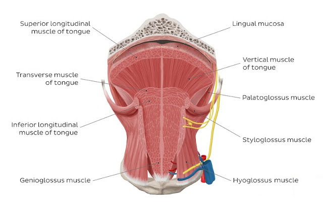
| Overview of tongue muscles (anterior view) |
The tongue is chiefly a muscular organ with some amount of fatty and fibrous tissue distributed throughout its substance. All the muscles of the tongue are paired structures, with each copy being found on either side of the median fibrous septum. There are muscles that extend outside of the organ to anchor it to surrounding bony structures, known as extrinsic muscles. The other set of muscles are confined to each half of the organ and contribute to altering the shape of the organ; these are the intrinsic muscles.
The intrinsic tongue muscles are responsible for adjusting the shape and orientation of the organ. It is made up of four paired muscles, which are discussed below in a dorsoventral manner.
The superior longitudinal muscles are made up of a thin layer of muscle fibers traveling in a mixture of oblique and longitudinal axes just deep to the superior mucosal surface of the organ. These fibers arise from the median fibrous septum as well as the fibrous layer of submucosa from the level of the epiglottis. They eventually insert along the lateral and apical margins of the organ. These muscles are responsible for retracting and broadening the tongue, as well as elevating the tip of the tongue. The net effect of these muscles results in shortening of the organ.
Another set of muscles occupy the dorsoventral plane of the tongue deep to the superior longitudinal muscles. These are the vertical muscles that arise from the root of the organ and genioglossus muscle and insert into the median fibrous septum, along the entire length of the organ. These muscles facilitate flattening and widening of the tongue.
References
No comments:
Post a Comment