Pages

Search This Blog

Wednesday, March 3, 2021

Vomiting

 The protective response of vomiting is an example of central regulation of gut motility functions. Vomiting starts with salivation and the sensation of nausea. Reverse peristalsis empties material from the upper part of the small intestine into the stomach. The glottis closes, preventing aspiration of vomitus into the trachea. The breath is held in mid inspiration. The muscles of the abdominal wall contract, and because the chest is held in a fixed position, the contraction increases intra-abdominal pressure. The LES and the esophagus relax, and the gastric contents are ejected. The “vomiting center” in the reticular formation of the medulla consists of various scattered groups of neurons in this region that control the different components of the vomiting act.

Neural pathways leading to the initiation of vomiting in response to various stimuli.


  Irritation of the mucosa of the upper gastrointestinal tract is one trigger for vomiting. Impulses are relayed from the mucosa to the medulla over visceral afferent pathways in the sympathetic nerves and vagi. Other causes of vomiting can arise centrally. For example, afferents from the vestibular nuclei mediate the nausea and vomiting of motion sickness. Other afferents presumably reach the vomiting control areas from the diencephalon and limbic system, because emetic responses to emotionally charged stimuli also occur. 

📖 Rosen & Barkin’s 5-Minute Emergency Medicine Consult 

Insulin receptors

Insulin receptors are found on many different cells in the body, including cells in which insulin does not increase glucose uptake. The insulin receptor, which has a molecular weight of approximately 340,000, is a tetramer made up of two α and two β glycoprotein subunits. All these are synthesized on a single mRNA and then proteolytically separated and bound to each other by disulfide bonds. 

Insulin, IGF-I, and IGF-II receptors. Each hormone binds primarily to its own receptor, but insulin also binds to the IGF-I receptor, and IGF-I and IGF-II bind to all three. The purple boxes are intracellular tyrosine kinase domains. Note the marked similarity between the insulin receptor and the IGF-I receptor; also note the 15 repeat sequences in the extracellular portion of the IGF-II receptor.
  Insulin, IGF-I, and IGF-II receptors. Each hormone binds primarily to its own receptor, but insulin also binds to the IGF-I receptor, and IGF-I and IGF-II bind to all three. The purple boxes are intracellular tyrosine kinase domains. Note the marked similarity between the insulin receptor and the IGF-I receptor; also note the 15 repeat sequences in the extracellular portion of the IGF-II receptor.

The gene for the insulin receptor has 22 exons and in humans is located on chromosome 19. The α subunits bind insulin and are extracellular, whereas the β subunits span the membrane. The intracellular portions of the β subunits have tyrosine kinase activity. The α and β subunits are both glycosylated, with sugar residues extending into the interstitial fluid.

📖 Endocrine Secrets 

Morison’s pouch

Morison’s pouch is also known as the posterior right subhepatic space or hepatorenal fossa. It separates the liver from the right kidney and is not filled with any fluid under normal conditions. It is a potential space, meaning a space that can occur between two adjacent structures that are normally pressed together. The anterior boundary consists of the right hepatic lobe and gallbladder.

Morison’s pouch

Posteriorly, there is the right kidney, right adrenal gland, the second part of the duodenum, hepatic flexure, and pancreatic head. The transverse mesocolon lies inferiorly. The posterior right subhepatic space communicates with the right subphrenic space and right paracolic gutter.

📖 Gray’s Atlas of Anatomy 3th Edition 

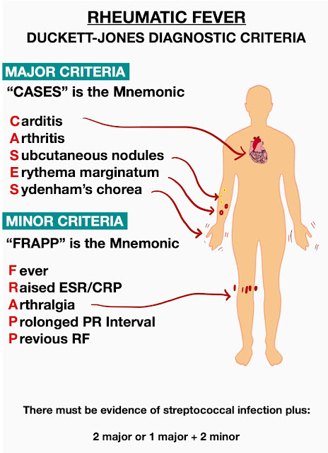
Jones criteria

The Jones Criteria for guidance in the diagnosis of acute rheumatic fever were first published by T. Duckett Jones, MD, in 1944 and have been revised over the years by the American Heart Association. The current guidelines are an update of these criteria.

The Jones criteria are used to diagnose rheumatic fever.

Jones criteria
Jones criteria 

The 5 major criteria consist of the following:
  • Carditis
  • Polyarthritis
  • Chorea
  • Erythema marginatum
  • Subcutaneous nodules
The minor criteria include the following:
  • Fever
  • Arthralgia
  • Elevated erythrocyte sedimentation rate or C-reactive protein level
  • Prolonged PR interval on electrocardiogram (ECG)
The presence of 2 major manifestations or of 1 major and 2 minor manifestations, supported by evidence of a preceding GAS infection by positive throat swab or culture results or by high serum ASO titers, strongly suggests ARF.

Epidermis

The outer layer, the epidermis, is relatively thin over most areas but is thickest on the palms of the hands and the soles of the feet. Although the epidermis is composed of several sublayers called strata, the  stratum corneum and the basal layer, which is the deepest layer, are of greatest importance.

Structure of the skin and subcutaneous tissue.

The stratum corneum is composed of dead, flat cells that lack a blood supply and sensory receptors. Its thickness is related to normal wear of the area it covers. 

📖 Ferri’s Fast Facts in Dermatology: A Practical Guide to Skin Diseases and Disorders