Caffey's landmark article of 1946 noted an association between healing long-bone fractures and chronic subdural hematomas in infancy, and it was the first to draw attention to physical abuse as a unifying etiology. In 1962, Caffey and Kempe et al proposed manhandling and violent shaking as mechanisms of injury and emphasized the acute and long-term sequelae of abuse as serious public health problems. Since these early reports, investigators have more clearly defined the pathophysiology of abusive injuries. Community-service and law-enforcement authorities have taken a role in protecting potential victims and in prosecuting perpetrators.
 |
| Caffey-Kempe syndrome |
In the United States, in 2015, there were 683,000 victims of child abuse, and approximately 1670 children died of abuse and neglect, a rate of 2.25 per 100,000 children. Almost 75% of those deaths occurred in children younger than 3 years. Most reports of abuse were submitted by educational personnel (18.4%) and legal and law enforcement personnel (18.2%). Approximatley 9% of reports were submitted by medical personnel.
For infants and children younger than 2 years, a skeletal survey should be performed as the initial screening examination when child abuse is being considered. The survey consists of the acquisition of a series of images collimated to each body region. The series includes frontal and lateral views of the skull, frontal and lateral views of the spine, frontal views of the chest (ribs) and pelvis, and frontal views of the extremities, including the hands and feet.
📖 Textbook of Paediatric Emergency Medicine 3rd Edition
The skeletal survey is widely available and inexpensive in comparison with alternative imaging modalities. Other important advantages of the skeletal survey include a high sensitivity for most acute and healing fractures and a relatively low radiation burden.
A babygram, in which the entire skeleton is depicted on a single image, is not an appropriate substitute for a properly performed survey. Geometric distortion and varying exposures are unacceptable limitations of this image. Use of a high-detail, high-contrast, screen-film system with good spatial resolution is mandatory. All abnormal areas should be viewed on at least 2 projections.
Computed tomography (CT) scanning of the head is the imaging modality of choice for evaluating a child with acute neurologic findings or retinal hemorrhage on physical examination. It is more sensitive to acute intracerebral and extra-axial hemorrhages than is magnetic resonance imaging (MRI). Brain MRI may be helpful as an adjunct for the evaluation of axonal shear injuries and for a precise dating of intracranial hemorrhage.
Subarachnoid hemorrhages (SAHs) are best demonstrated on CT scans. The use of MRI to detect acute SAH remains controversial. However, MRI is superior to CT scanning for differentiating a hypoattenuating subdural hematoma from cerebrospinal fluid (CSF) and for detecting small and chronic extra-axial fluid collections. (See the images below; both images reveal a subdural hematoma, the first with a CT scan and the second with an MRI scan.)
Rib fractures occur when a compressive force is applied simultaneously to the sternum and to the costovertebral junction during violent shaking as the perpetrator compresses the child's chest using both hands.
The posterior ribs are most commonly fractured, because the greatest force is imparted to the articulation of the head and to the neck of the rib with the transverse process of the vertebral body.
However, fractures are not limited to the posterior aspects of the ribs. Anterolateral fractures are also common. Rib fractures are typically noted at several contiguous levels; they are frequently bilateral.
 |
| Skull fracture secondary to child abuse horizontally crosses the left frontal region superior to the orbital rim. |
Head injury accounts for 80% of deaths associated with abuse in children younger than age 2 years. Mechanisms of injury include forceful shaking, either by itself or accompanied by abrupt impact.
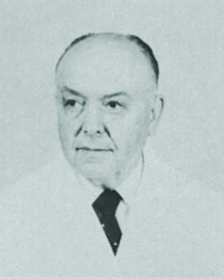
John Patrick Caffey (1895 - 1978), American paediatrician.
 |
| John Patrick Caffey |
John Patrick Caffey was born in 1895, the year Wilhelm Conrad Röntgen (1845-1923) discovered the x-rays. Caffey graduated in medicine at the University of Michigan in 1919, and served his internship at the Barnes Hospital, St. Louis. In 1920 he went to Warsaw in 1920 with the American Red Cross in Poland and Russia, but contracted typhus. Immense medical problems had arisen in these countries following World War I, and it was the lack of medical care for children that made him remain in Europe for three years after the convalescence, working with the Hoover Commission in Russia.
After returning to the USA, Caffey became chief resident at the University Hospital in Ann Arbor, Michigan. He then worked for a short period of time at the Babie’s Hospital in Columbia University, New York, before he went into private practice in New York. In 1929 he was appointed to the new Babie’s Hospital where he was assigned to take charge of radiology as the hospital's first in-house roentgenologist.
In 1950 he became professor of clinical paediatrics at the College of Physicians and Surgeons, Columbia University, New York, and four years later was appointed professor of radiology.
After his compulsory retirement in 1960 he became emeritus of radiology. Caffey had no intentions of leaving his work, however, and became visiting professor of paediatrics and radiology at the Children’s Hospital, University of Pittsburgh. He continued working until the day of his death in September 1978, at the age of 83 years.
Charles Henry Kempe (1922 - 1984), Poland paediatrician.
References
Caffey J. Multiple fractures in the long bones of infants suffering from chronic subdural hematoma. AJR Am J Roentgenol. 56:163-173.
Kempe CH, Silverman FN, Steele BF, Droegemueller W, Silver HK. The battered-child syndrome. JAMA. 1962 Jul 7. 181:17-24. [Medline].
U.S. Department of Health & Human Services. Child Maltreatment 2015. U.S. Department of Health & Human Services, Administration for Children and Families, Administration on Children, Youth and Families, Children’s Bureau. (2017). Available at http://www.acf.hhs.gov/programs/cb/research-data-technology/statistics-research/child-maltreatment. . 2017; Accessed: March 23, 2017.
Kleinman PK, Nimkin K, Spevak MR, et al. Follow-up skeletal surveys in suspected child abuse. AJR Am J Roentgenol. 1996 Oct. 167(4):893-6. [Medline].
Harper NS, Eddleman S, Lindberg DM. The utility of follow-up skeletal surveys in child abuse. Pediatrics. 2013 Mar. 131(3):e672-8. [Medline].
Kleinman PK, Morris NB, Makris J, Moles RL, Kleinman PL. Yield of radiographic skeletal surveys for detection of hand, foot, and spine fractures in suspected child abuse. AJR Am J Roentgenol. 2013 Mar. 200(3):641-4. [Medline].
van Rijn RR, Sieswerda-Hoogendoorn T. Educational paper: imaging child abuse: the bare bones. Eur J Pediatr. 2012 Feb. 171(2):215-24. [Medline]. [Full Text].
Kleinman PK, Marks SC. A regional approach to classic metaphyseal lesions in abused infants: the distal tibia. AJR Am J Roentgenol. 1996 May. 166(5):1207-12. [Medline].
Nimkin K, Kleinman PK. Imaging of child abuse. Radiol Clin North Am. 2001 Jul. 39(4):843-64. [Medline].
Pfeifer CM, Hammer MR, Mangona KL, Booth TN. Non-accidental trauma: the role of radiology. Emerg Radiol. 2017 Apr. 24 (2):207-213. [Medline].
Borg K, Hodes D. Guidelines for skeletal survey in young children with fractures. Arch Dis Child Educ Pract Ed. 2015 Oct. 100 (5):253-6. [Medline].
Greeley CS. Abusive head trauma: a review of the evidence base. AJR Am J Roentgenol. 2015 May. 204 (5):967-73. [Medline].
Vázquez E, Delgado I, Sánchez-Montañez A, Fábrega A, Cano P, Martín N. Imaging abusive head trauma: why use both computed tomography and magnetic resonance imaging?. Pediatr Radiol. 2014 Dec. 44 Suppl 4:S589-603. [Medline].
Kirks DR. Radiological evaluation of visceral injuries in the battered child syndrome. Pediatr Ann. 1983 Dec. 12(12):888-93. [Medline].
Lonergan GJ, Baker AM, Morey MK, Boos SC. From the archives of the AFIP. Child abuse: radiologic-pathologic correlation. Radiographics. 2003 Jul-Aug. 23(4):811-45. [Medline].
Rustamzadeh E, Truwit CL, Lam CH. Radiology of nonaccidental trauma. Neurosurg Clin N Am. 2002 Apr. 13(2):183-99. [Medline].
Greenberg MI, Hendrickson R, Silverberg M, et al, eds. Greenberg's Text-Atlas of Emergency Medicine. 2005.
Kemp AM, Dunstan F, Harrison S, Morris S, Mann M, Rolfe K, et al. Patterns of skeletal fractures in child abuse: systematic review. BMJ. 2008 Oct 2. 337:a1518. [Medline]. [Full Text].
Adams JA. Guidelines for medical care of children evaluated for suspected sexual abuse: an update for 2008. Curr Opin Obstet Gynecol. 2008 Oct. 20(5):435-41. [Medline].
Mok JY. Non-accidental injury in children--an update. Injury. 2008 Sep. 39(9):978-85. [Medline].
Kleinman PK, Blackbourne BD, Marks SC, et al. Radiologic contributions to the investigation and prosecution of cases of fatal infant abuse. N Engl J Med. 1989 Feb 23. 320(8):507-11. [Medline].
Slovis TL, Smith W, Kushner DC, et al. Imaging the child with suspected physical abuse. American College of Radiology. ACR Appropriateness Criteria. Radiology. 2000 Jun. 215 Suppl:805-9. [Medline].
Meyer JS, Gunderman R, Coley BD, Bulas D, Garber M, Karmazyn B, et al. ACR Appropriateness Criteria(®) on suspected physical abuse-child. J Am Coll Radiol. 2011 Feb. 8 (2):87-94. [Medline].
American College of Radiology. American College of Radiology ACR Appropriateness Criteria Suspected Physical Abuse--Child. American College of Radiology. Available at https://acsearch.acr.org/docs/69443/Narrative/. 2016; Accessed: March 23, 2017.
Paddock M, Sprigg A, Offiah AC. Imaging and reporting considerations for suspected physical abuse (non-accidental injury) in infants and young children. Part 1: initial considerations and appendicular skeleton. Clin Radiol. 2017 Mar. 72 (3):179-188. [Medline].
Paddock M, Sprigg A, Offiah AC. Imaging and reporting considerations for suspected physical abuse (non-accidental injury) in infants and young children. Part 2: axial skeleton and differential diagnoses. Clin Radiol. 2017 Mar. 72 (3):189-201. [Medline].
Paine CW, Fakeye O, Christian CW, Wood JN. Prevalence of Abuse Among Young Children With Rib Fractures: A Systematic Review. Pediatr Emerg Care. 2016 Oct 4. [Medline].
Conway JJ, Collins M, Tanz RR, et al. The role of bone scintigraphy in detecting child abuse. Semin Nucl Med. 1993 Oct. 23(4):321-33. [Medline].
Carty H. Non-accidental injury: a review of the radiology. Eur Radiol. 1997. 7(9):1365-76. [Medline].
Laor T, Jaramillo D. Metaphyseal abnormalities in children: pathophysiology and radiologic appearance. AJR Am J Roentgenol. 1993 Nov. 161(5):1029-36. [Medline].
Merten DF, Carpenter BL. Radiologic imaging of inflicted injury in the child abuse syndrome. Pediatr Clin North Am. 1990 Aug. 37(4):815-37. [Medline].
Dedouit F, Guilbeau-Frugier C, Capuani C, Sévely A, Joffre F, Rougé D, et al. Child Abuse: Practical Application of Autopsy, Radiological, and Microscopic Studies. J Forensic Sci. 2008 Aug 25. [Medline].
Makoroff KL, Cecil KM, Care M, Ball WS Jr. Elevated lactate as an early marker of brain injury in inflicted traumatic brain injury. Pediatr Radiol. 2005 Jul. 35(7):668-76. [Medline].
Child Welfare Information Gateway. Child Abuse and Neglect Fatalities: Statistics and Interventions. 2004. Updated July 24, 2006. Available at: http://www.childwelfare.gov/pubs/factsheets/fatality.cfm. [Full Text].


No comments:
Post a Comment