Pages

Search This Blog

Monday, March 22, 2021

Double inferior vena cava

Double inferior vena cava is a vein abnormality that is present from birth (congenital). Individuals with this anomaly have two inferior vena cavas instead of one. The inferior vena cava carries oxygen-poor blood from the lower parts of the body into the heart. Double inferior vena cava does not cause any symptoms. It is usually diagnosed when an imaging test, such as CT or MRI, is performed for other medical reasons. The reported incidence of double inferior vena cava ranges from 0.2 percent to 3 percent

Double inferior vena cava


A double or duplicated inferior vena cava results from lack of atrophy in part of the left supracardinal vein, resulting in a duplicate structure to the left of the aorta. The common form is a partial paired inferior vena cava that connects the left common iliac and left renal veins. When caval interruption, such as placement of a filter, is planned, these alternate pathways must be considered. As an alternative, the inferior vena cava may not develop. The most common alternate route for blood flow is through the azygous vein, which enlarges to compensate. If a venous stenosis is present at the communication of iliac veins and azygous vein, back pressure can result in insufficiency, stasis, or thrombosis.

📖 Netter’s Cardiology 2th Edition  

Fabry-Anderson disease

Anderson–Fabry disease (AFD) is a rare X-linked disorder caused by defects of the alpha-galactosidase (α-Gal) enzyme. Mutations in the Alpha Galactosidase gene (GLA), which encodes for α-Gal, affect synthesis, trafficking, folding, degradation and enzymatic activity of α-Gal resulting in progressive intracellular accumulation of globotriaosylceramide (Gb3). Intracellular Gb3 and related glycosphingolipids accumulation leads to organ/tissue damage potentially affecting cardiovascular, renal, gastrointestinal, cerebrovascular, neurologic, auditory, ocular and cutaneous systems. Recent evidence supports the hypothesis of a tissue-specific, mutation-dependent ‘affinity’ for Gb3 storage. The AFD clinical phenotype is characterized by variability in the age of onset and severity and can be severe and early in classic forms of AFD or mild and later in variant forms. Replicated evidence demonstrates that carriers of certain mutations in the GLA gene develop preferential, albeit nonexclusive, cardiac, renal and neurologic phenotypes: for example p.(Asn215Ser) and p.(Phe113Ile) are invariably associated with late-onset hypertrophic cardiomyopathy-like (HCM-like) phenotype.

Fabry-Anderson disease


Hemizygous males with the classic form of AFD demonstrate low or absent enzyme activity. The patients typically develop signs and symptoms in childhood or adolescence (delayed puberty and growth, gastrointestinal symptoms, corneal opacities, angiokeratomas, acroparesthesias/neuropathic pain).

Saturday, March 20, 2021

Galant's biceps reflex

Biceps reflex is a reflex test that examines the function of the C5 reflex arc and the C6 reflex arc. The test is performed by using a tendon hammer to quickly depress the biceps brachii tendon as it passes through the cubital fossa. Specifically, the test activates the stretch receptors inside the biceps brachii muscle which communicates mainly with the C5 spinal nerve and partially with the C6 spinal nerve to induce a reflex contraction of the biceps muscle and jerk of the forearm.

Galant's biceps reflex

A strong contraction indicates a 'brisk' reflex, and a weak or absent reflex is known as 'diminished'. Brisk or absent reflexes are used as clues to the location of neurological disease. Typically brisk reflexes are found in lesions of upper motor neurones, and absent or reduced reflexes are found in lower motor neurone lesions.

A change to the biceps reflex indicates pathology at the level of musculocutaneous nerve, segment C5/6 or at some point above it in the spinal cord or brain.

Left superior vena cava

Left superior vena cava (LSVC) is the most common congenital malformation of the thoracic venous return and is present in 0.3 to 0.5% of individuals in the general population with a normal heart, and 4.5% in individuals with congenital heart diseases. A LSVC co-occurs with the right superior vena cava in 80 to 90% of cases, 2 and may also be accompanied by other heart abnormalities, such as anomalous connections of the pulmonary veins, aortic coarctation, tetralogy of Fallot, transposition of the great vessels as well as dextroversion. Moreover, cardiac rhythm disturbances concerning impulse formation and conduction have been observed.

 
Left superior vena cava

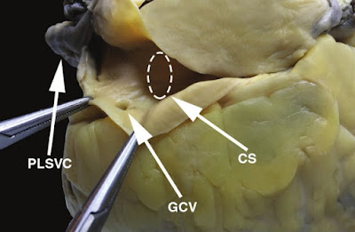
The LSVC usually drains into the right atrium (in 80–92%) through a dilated coronary sinus (CS),5,6 but in approximately 10 to 20% of cases, it is associated with left atrial (LA) drainage. The LSVC may drain directly through the left atrium or via the unroofed CS, which is a cause of right-to-left cardiac shunt. The majority of patients with LSVC are asymptomatic. In general, only patients with unusual drainage and right-to-left shunting are of clinical significance. Anomalous venous return via the LSVC may be the cause of cardiac arrhythmias, decreased exercise tolerance, progressive fatigue, chest discomfort, palpitations, syncope or cyanosis.

 
The left superior vena cava drains into the coronary sinus in this heart specimen. CS, coronary sinus; GCV, great cardiac vein; PLSVC, persistent left superior vena cava.

The implications of existing LSVC could be important for clinicians who are involved in placement of central venous-access devices.

📖 Netter’s Cardiology 2th Edition  

Thursday, March 18, 2021

Galant Reflex

 The Spinal Galant Reflex provides the opportunity to experience a change from whole body movements to same side (homolateral) movements. It emerges at 20 weeks in utero, is actively present at birth, and should be integrated by the time the baby is 3 to 9 months of age.

A deep abdominal reflex in newborn. There is a contraction of the abdominal muscles on tapping the anterior superior iliac spine or touching the skin along spine from shoulder to hip. In infancy it is physiologically present until about the sixth month. At higher ages it is always pathological.
The Spinal Galant Reflex should disappear by nine months, and retention beyond this point results in problems such as bed-wetting, fidgeting and the inability to sit still, short-term memory loss or an inability to concentrate.

The Spinal Galant Reflex is present when a baby is placed in a prone position (one their stomach) and stimulation is placed to one side of the spine. If the reflex is present, the hip on the side of the stimulus will flex (curl toward that side). This reflex helps the baby to work it’s way down the birth canal during the birthing process, and also allows the fetus to hear and feel the sound vibrations in the womb.

📖 Workbook in Practical Neonatology