Pages

Search This Blog

Tuesday, March 16, 2021

Eales' disease

 Eales Disease is a rare disorder of sight that appears as an inflammation and white haze around the outercoat of the veins in the retina. The disorder is most prevalent among young males and normally affects both eyes. Usually, vision is suddenly blurred because the clear jelly that fills the eyeball behind the lens of the eye seeps out (vitreous hemorrhaging).

Eales' disease
Eales' disease

Eales Disease usually presents as blurred vision resulting from oozing of the clear jelly-like substance from behind the lens of the eye. At the onset of the disorder, the small outer veins of the retina show sheathing (encapsulation or covering). As the disease progresses, the inflammation around the veins in the retina extends further behind the lens. Eales Disease may also be associated with peripheral retinal neovascularization which is the formation of new blood vessels on the outer part of the retina.

 📖 The Massachusetts Eye and Ear Infirmary Illustrated Manual of Ophthalmology 5th Edition 

Monday, March 15, 2021

Eagle’s syndrome

Eagle’s syndrome, defined by Eagle in 1949, is characterised by morphological abnormality/ossification of the styloid process. The average length of the styloid process is 20-30 mm in the adult Caucasians and 15.4-18.8 mm in the Asian population. An elongated styloid process is defined by being at least 30 mm long. The clinical picture is composed of recurrent throat and neck pain, radiating into the ear and dysphagia. Symptoms can be bilateral or, more frequently, unilateral. The incidence is 4-8 per 10,000 people.

Eagle’s syndrome
Eagle’s syndrome

The aetiology is not well defined and a number of theories have been suggested, such as congenital elongation due to the persistence of an embryonic cartilaginous outgrowth, calcification of the stylohyoid ligament and formation of bone tissue at the insertion of the ligament. This clinical picture might also be seen in patients after tonsillectomy.

📖 Merritt’s Neurology Handbook 10th Edition 

Dabska's tumour

 Papillary intralymphatic angioendothelioma (Dabska tumor) is a rarely metastasizing lymphatic vascular neoplasm that usually affects children and young adults. The majority of these cases occur in soft tissues of extremities, and to date less than 40 cases have been described. Despite the generally indolent evolution, can be locally invasive with the potential to metastasize. 

Dabska's tumour - Violaceous lesion, with poorly defined limits, on the sole of the right foot.

Papillary intralymphatic angioendothelioma (PILA or Dabska tumor) is a rare vascular neoplasm, defined as low grade malignancy tumor, with high tendency of local recurrence.

Histopathology. Papillary projections with vascular axes lined by rounded and hyperchromatic cells – “Hobnail cells” (Hematoxylin & eosin, ×100).

Originally considered a malignant tumor and called malignant endovascular papillary hemangioendothelioma, it was renamed in 1998, due to its borderline behavior, prominent presence of lymphatic vessels and the presence of intravascular papillary proliferation. 

📖 Diagnostic Pathology: Soft Tissue Tumors 3rd Edition 

Caffey-Kempe syndrome

 Caffey's landmark article of 1946 noted an association between healing long-bone fractures and chronic subdural hematomas in infancy, and it was the first to draw attention to physical abuse as a unifying etiology. In 1962, Caffey and Kempe et al proposed manhandling and violent shaking as mechanisms of injury and emphasized the acute and long-term sequelae of abuse as serious public health problems. Since these early reports, investigators have more clearly defined the pathophysiology of abusive injuries. Community-service and law-enforcement authorities have taken a role in protecting potential victims and in prosecuting perpetrators.

Caffey-Kempe syndrome

In the United States, in 2015, there were 683,000 victims of child abuse, and approximately 1670 children died of abuse and neglect, a rate of 2.25 per 100,000 children. Almost 75% of those deaths occurred in children younger than 3 years. Most reports of abuse were submitted by educational personnel (18.4%) and legal and law enforcement personnel (18.2%). Approximatley 9% of reports were submitted by medical personnel.

For infants and children younger than 2 years, a skeletal survey should be performed as the initial screening examination when child abuse is being considered. The survey consists of the acquisition of a series of images collimated to each body region. The series includes frontal and lateral views of the skull, frontal and lateral views of the spine, frontal views of the chest (ribs) and pelvis, and frontal views of the extremities, including the hands and feet. 

📖 Textbook of Paediatric Emergency Medicine 3rd Edition 

Sunday, March 14, 2021

Babington’s disease

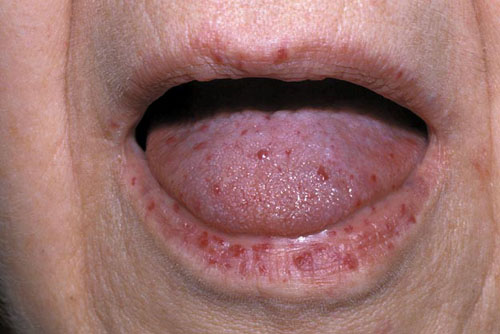
Babington’s disease  - Osler-Weber-Rendu syndrome (OWR) is also known as hereditary hemorrhagic telangiectasia (HHT). It’s a genetic blood vessel disorder that often leads to excessive bleeding. According to the HHT Foundation International, the syndrome affects about one in 5,000 people. However, many people with the disease don’t know they have it, so this number may actually be higher.

Babington’s disease
Babington’s disease 

The name Osler-Weber-Rendu syndrome is named for the doctors who worked on researching this condition in the 1890s. They discovered that issues with blood clotting don’t cause this condition, which was previously assumed. Instead, this condition is caused by problems with the blood vessels themselves.

Hereditary hemorrhagic telangiectasia (Osler-Weber-Rendu syndrome) is an autosomal dominant disorder that manifests with widespread cutaneous, mucosal, and visceral telangiectases and arteriovenous malformations. Papular, punctate, and linear telangiectases occur predominantly on the tongue, lips, digit tips, perioral region, and trunk.
Babington’s disease