Pages

Search This Blog

Sunday, March 14, 2021

Babcock's operation

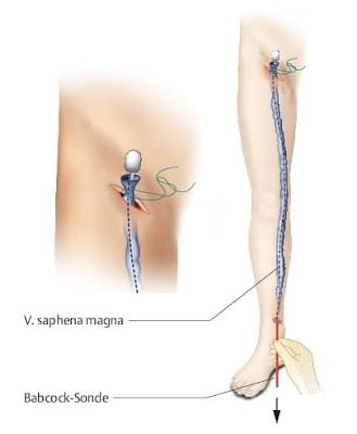
 The affected veins in the superficial venous system are pulled out through small incisions in the skin (stripped). This procedure is also known as "Babcock's procedure" or "Babcock's stripping". The American doctor Babcock described the method for the first time in 1907. This classic method is used to remove the affected segments of the worst affected long saphenous vein that runs from the ankle up to the inguinal fold.

Babcock's operation
Babcock's operation

The saphenous vein can only be removed, if the deep venous system is healthy. This is first checked by an ultrasound examination (Doppler ultrasonography), sometimes also with contrast medium X-rays (phlebography). Several days before the procedure, patients should not drink any alcohol or take medicines that delay blood clotting. These include most painkillers.

📖 Clinical Surgery: With Student Consult Access 

Friday, March 12, 2021

Pelvic plexus

The pelvic plexus is an association of neurons that govern visceral tissues involved in eliminative and reproductive functions. 
Pelvic plexus
Pelvic pleuses


It is the singular site in the autonomic nervous system where sympathetic and parasympathetic neurons occur in the same ganglia. Within the plexus, ganglia are not randomly positioned; sympathetic neurons tend to occur more ventrally while parasympathetic neurons are located more dorsally, both in accordance with the location of their target tissues and the entry point of their corresponding preganglionic nerve tracts. For example, the vas deferens and seminal vesicle are ventral in position and thus are innervated by more ventrally located pelvic neurons. Neurochemical studies of pelvic ganglia indicate that there are some characteristic associations of putative neurotransmitters which are based on target organ distribution and in part, dictated by the variety of target tissues within each organ. Penile neurons comprise a uniform population in that they are cholinergic and also may release vasoactive intestinal polypeptide (VIP) and nitric oxide. In contrast, target tissues of the internal genitalia are more diverse, requiring adrenergic and nonadrenergic innervation and a complementary neuropeptide. Preganglionic innervation may also be coded and although sympathetic and parasympathetic fibers are cholinergic, they may differ in respect to neuropeptides and nitric oxide. Sensory neuron collaterals may also influence principal neurons as do intrinsic neurons such as small intensely fluorescent cells. Transmission through pelvic ganglia may be simple as is apparent in penile innervation, or shows a greater integrative capacity, as exemplified by the innervation of the urinary bladder. The extent of interaction of sympathetic and parasympathetic pathways at the level of the pelvic plexus remains largely unknown.

Calciphylaxis

Calciphylaxis (calcific uremic arteriolopathy) results from deposition of calcium in the tunica media portion of the small vessel walls in association with proliferation of the intimal layer of endothelial cells. 

Calciphylaxis

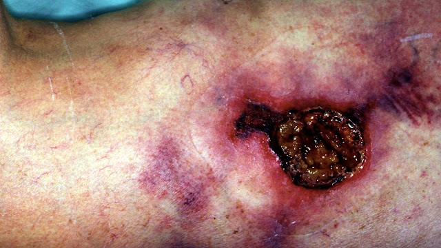
It is almost always associated with end-stage renal disease, especially in patients undergoing chronic dialysis (either peritoneal dialysis or hemodialysis). It has been reported to occur in up to 5% of patients who have been on dialysis for longer than 1 year.  Calciphylaxis typically manifests as nonhealing skin ulcers located in adiposerich areas of the trunk and thighs, but the lesions can occur anywhere. They are believed to be caused by an abnormal ratio of calcium and phosphorus, which leads to the abnormal deposition within the tunica media of small blood vessels. This eventually results in thrombosis and ulceration of the overlying skin. Calciphylaxis has a poor prognosis, and there are few well-studied therapies. 

Clinical Findings: Calciphylaxis is almost exclusively seen in patients with chronic end-stage renal disease. Most patients have been on one form of dialysis for at least 1 year by the time of presentation. 

Calciphylaxis
Calciphylaxis

The initial presenting sign is that of a tender, dusky red to purple macule that quickly ulcerates. The ulcerations have a ragged border and a thick black necrotic eschar. The ulcers tend to increase in size, and new areas appear before older ulcers have any opportunity to heal. 

📖 Critical Care Nephrology 3rd Edition 

Wednesday, March 10, 2021

Dagnini’s reflex

Dagnini’s reflex ( Aschner-Dagnini test, Oculocardiac Reflex)

Dagnini’s reflex ( Aschner-Dagnini test, Oculocardiac Reflex )

The oculocardiac reflex (OCR), also known as the Aschner reflex or trigeminovagal reflex (TVR), was first described in 1908 as a reduction in heart rate secondary to direct pressure placed on the eyeball. It is defined by a decrease in heart rate by greater than 20% following globe pressure or traction of the extraocular muscles. The reflex most commonly results in sinus bradycardia. However, it also has a reported association with reduced arterial pressure, arrhythmia, asystole, and even cardiac arrest. This reflex has most notably been depicted during ophthalmologic procedures, more specifically during strabismus surgery; however, it also may be activated by facial trauma, regional anesthetic nerve blocks, and mechanical stimulation. This activity describes the oculocardiac reflex and highlights the role of the interprofessional team in the management of patients with the symptoms during surgery.

Anatomy and Physiology

The OCR arc consists of an afferent and efferent limb. The trigeminal nerve, otherwise known as the fifth cranial nerve, serves as the sensory afferent limb.

📖 Hurst’s the Heart, 14th Edition: Two Volume Set 

Baastrup's sign

(Baastrup’s disease, Kissing spines syndrome)

Excessive lordosis is a common finding and may produce mechanical pressure that causes repetitive strains of the interspinous ligament with subsequent degeneration and collapse. Baastrup’s disease (kissing spine syndrome) is a term referring to close approximation of adjacent spinous processes due to degenerative changes of the spine. 

Baastrup’s disease
Baastrup’s disease

Baastrup’s disease usually affects the lumbar spine, with L4-L5 being the most commonly affected level. There is higher occurrence at ages over 70 and no gender predilection. Symptoms include back pain with midline distribution that worsens during extension, is relieved during flexion and is exaggerated upon finger pressure at the level of interest. Diagnosis rests on clinical examination and imaging studies. The hallmark of imaging findings is the close approximation and contact of adjacent spinous processes, with all the subsequent findings including oedema, cystic lesions, sclerosis, flattening and enlargement of the articulating surfaces, bursitis and occasionally epidural cysts or midline epidural fibrotic masses. Proposed therapies include conservative treatment, percutaneous infiltrations or surgical therapies such as excision of the bursa or osteotomy. 

Pathogenesis

Baastrup’s disease is a term referring to close approximation of adjacent spinous processes due to general degenerative changes of the spine. Usually there is an excessive lordosis with resultant mechanical pressure process that causes repetitive strains of the interspinous ligament with subsequent degeneration and collapse.

📖 Practical Office Orthopedics